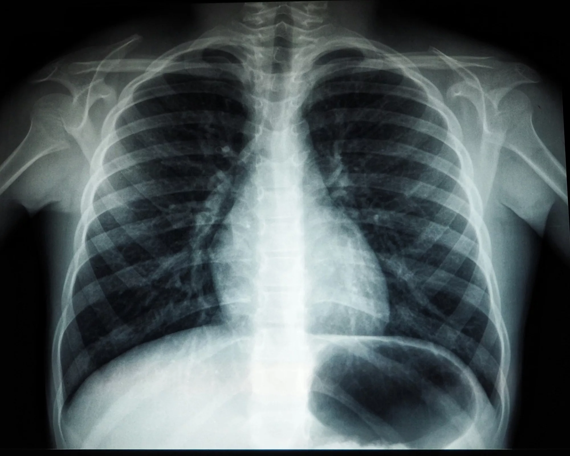
In the world of medical imaging, radiography and sonography stand as two of the most commonly utilized technologies, each serving unique purposes in diagnosing and treating a wide range of conditions. Both play a critical role in modern healthcare by enabling physicians to visualize the inside of the human body without the need for invasive procedures. However, while radiography and sonography share the goal of improving diagnostic accuracy and patient outcomes, the methods they use, the principles they are based on, and the applications they serve are vastly different.
Radiography, often associated with X-rays, relies on ionizing radiation to create images of the body’s internal structures. It is particularly effective for visualizing bones, detecting fractures, and identifying abnormalities in the chest or abdomen. On the other hand, sonography, also known as ultrasound, uses high-frequency sound waves to produce real-time images, making it invaluable for examining soft tissues, monitoring pregnancies, and guiding certain medical procedures.
This article explores the fundamental differences between radiography and sonography, examining their underlying technologies, procedures, applications, advantages, and limitations. By understanding the distinctions between these imaging modalities, we can better appreciate their contributions to healthcare and how they complement each other in the diagnosis and management of diseases.
The Basics of Radiography
What Is Radiography?
Radiography is a medical imaging technique that uses ionizing radiation, primarily in the form of X-rays, to create images of the body’s internal structures. When X-rays are directed at the body, they pass through tissues at varying degrees depending on the tissue’s density. Dense materials like bones absorb more X-rays and appear white on the resulting image, while less dense tissues like muscles or fat allow more X-rays to pass through, appearing in shades of gray.
Radiography is one of the oldest and most widely used imaging techniques, dating back to its discovery by Wilhelm Conrad Roentgen in 1895. Over the years, it has evolved to include a variety of specialized applications, such as fluoroscopy and computed tomography (CT).
How Does Radiography Work?
Radiography involves the following components and steps:
- X-Ray Source: A machine generates X-rays and directs them toward the part of the body being examined.
- Patient Positioning: The patient is carefully positioned to ensure the X-rays target the correct area.
- Image Capture Device: As X-rays pass through the body, they are captured by a detector or film placed on the opposite side of the patient. This device records the pattern of X-ray absorption, producing an image.
The resulting images are called radiographs and provide a two-dimensional view of the body’s internal structures.
Common Uses of Radiography
Radiography is a versatile imaging tool used in various medical specialties. Some common applications include:
- Bone Imaging: Detecting fractures, dislocations, or bone deformities.
- Chest X-Rays: Diagnosing conditions like pneumonia, tuberculosis, or lung cancer.
- Abdominal Imaging: Identifying blockages, foreign objects, or gastrointestinal perforations.
- Dental Radiography: Visualizing teeth, jawbones, and oral health issues.
Advantages of Radiography
Radiography offers several benefits:
- Quick and Efficient: Radiographic exams typically take only a few minutes to perform.
- Widely Available: Most healthcare facilities have X-ray equipment.
- High-Resolution Bone Imaging: Ideal for diagnosing fractures and skeletal conditions.
Limitations of Radiography
Despite its advantages, radiography has limitations:
- Radiation Exposure: Ionizing radiation can be harmful in large doses, requiring careful monitoring and protective measures.
- Limited Soft Tissue Visualization: Radiography is less effective at imaging soft tissues like muscles or organs compared to other modalities.
The Basics of Sonography
What Is Sonography?
Sonography, also known as ultrasound imaging, is a medical imaging technique that uses high-frequency sound waves to produce real-time images of the body. Unlike radiography, which relies on radiation, sonography is based on sound waves that are emitted and reflected by different tissues within the body.
Sonography is particularly effective for imaging soft tissues, such as muscles, blood vessels, and organs. Its ability to produce dynamic, real-time images makes it a preferred choice for monitoring pregnancies, guiding needle biopsies, and assessing blood flow.
How Does Sonography Work?
Sonography involves the following components and steps:
- Transducer: A handheld device called a transducer emits high-frequency sound waves into the body.
- Sound Wave Reflection: As sound waves encounter different tissues, they are reflected back to the transducer at varying speeds and intensities.
- Image Formation: The transducer converts the reflected sound waves into electrical signals, which are processed by a computer to create real-time images.
Sonography produces two-dimensional, three-dimensional, or even four-dimensional images, depending on the equipment and application.
Common Uses of Sonography
Sonography is a highly adaptable imaging tool used in many medical fields. Common applications include:
- Obstetrics and Gynecology: Monitoring fetal development, diagnosing ectopic pregnancies, and assessing reproductive organs.
- Cardiology: Evaluating heart function through echocardiography.
- Abdominal Imaging: Visualizing organs like the liver, kidneys, and gallbladder.
- Vascular Imaging: Assessing blood flow and detecting blockages or clots.
Advantages of Sonography
Sonography has several notable advantages:
- No Radiation: Unlike radiography, ultrasound imaging does not use ionizing radiation, making it safer for patients, particularly pregnant women and children.
- Real-Time Imaging: Allows for dynamic assessments, such as observing fetal movement or blood flow.
- Portable and Accessible: Many ultrasound machines are compact and can be used in various settings, including bedside exams.
Limitations of Sonography
Despite its versatility, sonography has limitations:
- Limited Penetration: High-frequency sound waves struggle to penetrate dense tissues or bones, making it unsuitable for imaging the skeleton.
- Operator Dependence: Image quality and accuracy depend heavily on the skill and experience of the technician or physician performing the exam.
- Artifacts: Air and gas can interfere with sound wave transmission, making it difficult to image certain areas, such as the intestines.
Key Differences Between Radiography and Sonography
1. Underlying Technology
- Radiography: Uses ionizing radiation (X-rays) to create images.
- Sonography: Uses high-frequency sound waves to produce images.
The fundamental difference lies in the type of energy each technique uses—radiography relies on radiation, while sonography utilizes sound.
2. Imaging Capabilities
- Radiography: Excels at imaging dense structures like bones and detecting calcifications or foreign objects.
- Sonography: Specializes in visualizing soft tissues and real-time movement, such as fetal development or blood flow.
3. Safety Profile
- Radiography: Involves exposure to ionizing radiation, which must be minimized to reduce the risk of long-term harm.
- Sonography: Does not involve radiation and is considered safe for all patient populations.
4. Accessibility and Portability
- Radiography: Typically requires stationary equipment found in dedicated imaging suites.
- Sonography: Can be performed using portable machines, making it more accessible in outpatient or emergency settings.
5. Applications
- Radiography: Primarily used for skeletal imaging, chest X-rays, and dental assessments.
- Sonography: Commonly used for obstetrics, abdominal imaging, and vascular studies.
Complementary Roles in Healthcare
Although radiography and sonography differ significantly, they often complement each other in clinical practice. For example:
- Emergency Medicine: Radiography is used to identify fractures, while sonography can detect internal bleeding or organ damage.
- Cancer Diagnosis: Radiography may reveal tumors or masses, while sonography can guide biopsies for more accurate sampling.
- Cardiology: X-rays may assess the size of the heart, while echocardiography evaluates its function.
By combining these imaging modalities, healthcare providers can obtain a more comprehensive understanding of a patient’s condition.
Emerging Technologies and Future Trends
Both radiography and sonography are evolving rapidly, driven by advancements in technology and a growing emphasis on patient safety and diagnostic precision.
Advances in Radiography
- Digital Radiography: Offers faster image acquisition and improved resolution compared to traditional film-based methods.
- Low-Dose Imaging: Reduces radiation exposure without compromising image quality.
- 3D and 4D Imaging: Enhances the ability to visualize complex structures in three or four dimensions.
Advances in Sonography
- Elastography: Measures tissue stiffness, aiding in the diagnosis of liver fibrosis or tumors.
- Portable Ultrasound Devices: Increasingly compact and user-friendly machines make sonography more accessible in remote or underserved areas.
- Artificial Intelligence (AI): Improves image interpretation and assists technicians in real-time diagnostics.
Conclusion
Radiography and sonography are two distinct yet complementary imaging modalities that play vital roles in modern medicine. Radiography’s strength lies in its ability to capture detailed images of bones and dense structures, while sonography excels at visualizing soft tissues and dynamic processes without the risks associated with radiation.
By understanding the differences between these techniques, healthcare professionals can choose the most appropriate imaging modality for each clinical scenario, ensuring accurate diagnoses and optimal patient care. As technology continues to advance, both radiography and sonography will undoubtedly become even more powerful tools in the ever-evolving field of medical imaging.


